Labelled Diagram Of Onion Cell
Onion cell epidermal diagram labeled cells microscope under drawing skin epidermis lab bulb mag membrane observation vacuole nucleus leaves preparation Onion magnification 400x 100x Onion skin 200x plant slides dissection
Onion Cells under Microscope - Saurabh Garg
Onion cell diagram drawing Draw the figure of an onion peel showing cell Onion cell 400x lab microscope under labeled cells structure scoop science looked
Onion cell peel draw cytoplasm membrane vacuole showing brainly figure
Cells deixa comentariOnion epidermal cell labeled diagram Onion cells microscope blue methylene stained under observation umberto flickrOnion skin 200x « dissection connection.
Onion_cells – biobiznewsOnion cells under a microscope Onion cells microscope under magnified times cell 100x does genetics wallOnion cells at 400x magnification.

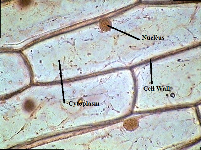
The science scoop: onion cell lab
Onion cells under microscopeOnion cells cell skin cytoplasm nucleolus vacuole nucleus wall strands principles biology ppt powerpoint presentation .
.


